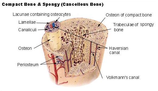
Compact Bone Diagram : Compact Bone Spongy Bone And Other Bone Components Human Anatomy And Physiology Lab Bsb 141. The diagram above shows a longitudinal view of an osteon. Compact and spongy.the names imply that the two types differ in density, or how tightly the tissue is packed together. (b) in this micrograph of the osteon, you can clearly see the concentric lamellae and central canals. Compact bone is formed in concentric circles. They allow blood vessels and nerves to travel through them to supply the osteocytes.
There are two types of bone tissue: The cells of compact bone, which is also called cortical bone, appear to be tightly packed into a solid mass. The main type of bone cell is the osteocyte (bone cell, shown as purple in the diagram). Haversian canals (sometimes canals of havers) are a series of microscopic tubes in the outermost region of bone called cortical bone. Compact bone structure diagram quizlet from o.quizlet.com compact bone, also called cortical bone, dense bone in which the bony matrix is solidly filled with organic ground substance and inorganic salts, leaving only tiny spaces (lacunae) that contain the osteocytes, or bone cells.compact bone makes up 80 percent of the human skeleton;
Anatomy Microscopic Structure Of Compact Bone Diagram Part 1 Diagram Quizlet from o.quizlet.com There are two types of bone tissue: Bone lamellae are arranged in regular haversian system. Online quiz to learn compact bone diagram; About press copyright contact us creators advertise developers terms privacy policy & safety how youtube works test new features press copyright contact us creators. The endosteum can be seen in the t.s. Spongy bones fills the inner layers of most of the bones. The two main structural components typically include spongy bone on the interior, with an outer layer of compact bone. The cells of compact bone, which is also called cortical bone, appear to be tightly packed into a solid mass.
Compact bone, also called cortical bone, is the hard, stiff, smooth, thin, white bone tissue that surrounds all bones in the human body.
Compact bone is formed in concentric circles. The outer part of a long bone is made of compact bone. (b) in this micrograph of the osteon, you can clearly see the concentric lamellae and central canals. Bone lamellae are arranged as interlocking networks. Add to favorites 0 favs. Long bones — a subtype of bones — are longer than they are wide. Have you ever seen fossil remains of dinosaur. If the outer layer of a cranial bone fractures, the brain is still protected by the intact inner layer. It makes up the outer cortex of all bones and is in immediate contact with the periosteum. Compact bone, also called cortical bone, dense bone in which the bony matrix is solidly filled with organic ground substance and inorganic salts, leaving only tiny spaces (lacunae) that contain the osteocytes, or bone cells.compact bone makes up 80 percent of the human skeleton; Microscopic structures of compact bone wedge of bone duration. Choose from 500 different sets of flashcards about long bone diagram on quizlet. 33 label the bone model these pictures of this page are about:compact bone labeled diagram compact bone diagram osteon compact bone ap pinterest anatomy human anatomy and.
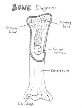
13 photos of the compact bone diagram labeled. Long bones — a subtype of bones — are longer than they are wide. You need to get 100% to score the 15 points available. Diagram of a typical long bone showing both cortical (compact) and cancellous (spongy) bone. The outer part of a long bone is made of compact bone.
Seer Training Structure Of Bone Tissue from training.seer.cancer.gov The main type of bone cell is the osteocyte (bone cell, shown as purple in the diagram). Diagram of a typical long bone showing both cortical (compact) and cancellous (spongy) bone. The cells of compact bone, which is also called cortical bone, appear to be tightly packed into a solid mass. Cortical bone is compact bone while cancellous bone is trabecular and spongy bone. Flat bones, like those of the cranium, consist of a layer of diploë (spongy bone), lined on either side by a layer of compact bone (). Illustration about compact bone, also called cortical bone, is the hard, stiff, smooth, thin, white bone tissue that surrounds all bones in the human body. (b) in this micrograph of the osteon, you can clearly see the concentric lamellae and central canals. Compact bone structure diagram quizlet from o.quizlet.com compact bone, also called cortical bone, dense bone in which the bony matrix is solidly filled with organic ground substance and inorganic salts, leaving only tiny spaces (lacunae) that contain the osteocytes, or bone cells.compact bone makes up 80 percent of the human skeleton;
If the outer layer of a cranial bone fractures, the brain is still protected by the intact inner layer.
Human bone generally comprises osseous tissue, an outer coating called a periosteum, and bone marrow. Compact bone is formed from a number of osteons, which are circular units of bone material and blood vessels. The main type of bone cell is the osteocyte (bone cell, shown as purple in the diagram). Microscopic structures of compact bone wedge of bone duration. It makes up the outer cortex of all bones and is in immediate contact with the periosteum. Compact and spongy.the names imply that the two types differ in density, or how tightly the tissue is packed together. Add to favorites 0 favs. Compact bone diagram bone cross section diagram file624 diagram of compact bone new. Under periosteum of all bones is the bulk of the diaphysis of long bones. The two layers of compact bone and the interior spongy bone work together to protect the internal organs. There are pores and spaces even in compact bone. The diagram above shows a longitudinal view of an osteon. Compact bone is formed in concentric circles.
(b) in this micrograph of the osteon, you can clearly see the concentric lamellae and central canals. Microscopic structures of compact bone wedge of bone duration. The two main structural components typically include spongy bone on the interior, with an outer layer of compact bone. The two layers of compact bone and the interior spongy bone work together to protect the internal organs. The endosteum can be seen in the t.s.
Simple Bone Diagram By Tessa Arnett Teachers Pay Teachers from ecdn.teacherspayteachers.com Touch device users can explore by touch or with. As seen in the image below, compact bone forms the cortex, or hard outer shell of most bones in the body. About press copyright contact us creators advertise developers terms privacy policy & safety how youtube works test new features press copyright contact us creators. Compact bone diagram osteon compact bone ap pinterest anatomy human anatomy and. Choose from 500 different sets of flashcards about long bone diagram on quizlet. Diagram of a typical long bone showing both cortical (compact) and cancellous (spongy) bone. They allow blood vessels and nerves to travel through them to supply the osteocytes. Bone marrow diagram, compact bone diagram quiz, compact bone slide labeled, diagram long bone, labeled compact bone model, human anatomy, bone marrow diagram, compact bone diagram quiz, compact bone slide labeled, diagram long bone, labeled compact bone model.
(b) in this micrograph of the osteon, you can clearly see the concentric lamellae and central canals.
Compact bone is formed from a number of osteons, which are circular units of bone material and blood vessels. It is also called osseous tissue or cortical bone and it provides structure and support for an organism as part of its skeleton, in addition to being a location for the storage of minerals like calcium.about 80% of the weight of the human skeleton comes from. As seen in the image below, compact bone forms the cortex, or hard outer shell of most bones in the body. The two main structural components typically include spongy bone on the interior, with an outer layer of compact bone. (b) in this micrograph of the osteon, you can clearly see the concentric lamellae and central canals. It makes up the outer cortex of all bones and is in immediate contact with the periosteum. Microscopic structures of compact bone wedge of bone duration. Compact bone is the strongest form of bone tissue containing few spaces. About press copyright contact us creators advertise developers terms privacy policy & safety how youtube works test new features press copyright contact us creators. The remainder of the bone is formed by cancellous or spongy bone. Compact bone diagram bone cross section diagram file624 diagram of compact bone new. Bone lamellae are arranged in regular haversian system. Illustration about compact bone, also called cortical bone, is the hard, stiff, smooth, thin, white bone tissue that surrounds all bones in the human body.